|
|

Step 3:
2 long axis views through the middle of the LV. Overlap if possible. Use slightly smaller PEG size in order to increase # of cardiac phases which will improve image quality making it easier to determine the short axis orientation
 |
Step 4:
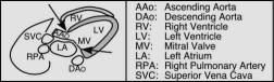
3 short axis slices, one below the valve plane (in the atria), one just above the valve plane (large LV and RV views) and one between the mid ventricle and apex. These images can be used to determine the optimal FOV and phase sampling ratio to prevent aliasing. Sometimes it is hard to decide on the exact angle to use. The simplest way to obtain consistent results is to locate the base of the LV wall that defines the valve planes as pointed out by the arrows in the previous diagram. Prescribe your Short Axis views parallel to this plane. Please note: this step (acquiring 3 SA views) can be skipped if you are already confident that you have adequate FOV and PSR values.
 |
Step 5 Short Axis:
Acquire all the short axis images needed from below the base (in the atria) to the apex using 8 to 10 mm thick slices, 12 to 16 cardiac phases which usually means a PEG size of 3 to 6, and the smallest FOV possible to prevent aliasing. The prescription angle is defined as above in step 4.